Trauma

Preserving our Urban Paradise

Written by: Samantha Martinez, Environmental Educator

Barn Owl 22-842

In Miami, we are constantly growing and upgrading our infrastructure as new homes, highways, and businesses are built daily to expand our urban paradise. This, on occasion, can come at the cost of the homes of our wild neighbors. This is what may have happened to patient 21-842, a juvenile Barn Owl. This owl was found on a construction site after a tree, believed to be harboring the bird's nest, was cut down. Unable to fly and with dry blood covering his feet and nares, the owl was brought into Pelican Harbor Seabird Station.  We see this all too often at PHSS, especially during the spring months as people prepare for hurricane season.

Once in care, our rehabbers noticed swelling and lameness in this patient's right leg, with x-rays confirming a midshaft fracture to the tarsometatarsus bone. The rehab team thoroughly cleaned the area and added a half splint with firm cast material to immobilize the young owl's leg during the healing process. This, coupled with supportive care, antibiotics, and pain medication, has this bird on the road to recovery. However, there is still a long way to go, and not all displaced fledglings are so lucky.

Wildlife rehabber, Carolina, renesting a displaced Eastern Screech Owl nestling.

We understand that tree trimming and construction are often unavoidable facts of urban life, especially considering the need to prevent damage from hurricanes. However, we recommend some simple ways to help avoid harm to wildlife when starting a building project or trimming trees for whatever reason. Such methods include doing an initial survey of the area to see if any wildlife is inhabiting it. You can check for droppings, nests, or nesting materials, and listen for animals in the immediate vicinity. If you find that an animal is nesting in a tree you plan to trim, we recommend leaving all branches supporting the nest as well as those protecting it from weather and predators until all babies have left the nest. Most birds will be done nesting by mid-July. Following these simple steps will help save countless nesting birds and mammals and prevent you from receiving costly fines associated with disturbing a federally protected nest. To learn more about protecting wildlife while trimming trees, feel free to reach out to us for a copy of our Animal Safety for Arborist sheet. 

Saved by the Power of Light

Written by: Milena Orjuela, PHSS Intern

Pelicans are meant to glide through the sky, dipping in and out of the ocean on a sunny day, and leaving your jaw dropped if they happen to catch a fish near you. That is what I thought until Pelican 21-372 came to our clinic. After taking a quick glance at the bird, I secretly thought to myself, “you are doomed.” Pelican 21-372 first came into our care after being spotted with monofilament entangling its left wing, upper body, and head. It was obvious upon examination that the bird was also malnourished and dehydrated. Our staff noted multiple open wounds in its face, abdomen, legs, and a large malodorous wound on the left patagium. A malodorous wound is characterized by necrotic tissue, or bacterial colonization, often containing both anaerobes and aerobes and is a major obstruction to healing. The patagium, the site of the open wound, is a web of elastic fibers that extends from the head to the wrist and has the very important job of assisting birds in gliding and flying. Such a grave injury on a crucial part of the bird’s wing is why Pelican 21-372’s chances were initially seen as quite poor. 

A set of X-rays determined that there were no fractures or broken bones to deal with in addition to the obvious open wounds. Subcutaneous fluids were given to aid with dehydration, antibiotics, pain killers, anti-inflammatories, and Vitamin E were administered to help the healing process, and the pelican was given one dose of anti-parasitic medication. All wounds were thoroughly cleansed and special care was given to the left wing. All dead tissue was removed from the patagium, a topical antibiotic ointment was administered, and the wing was wrapped. Though the physical treatments and medications certainly had a very beneficial role in healing the bird, the laser therapy Pelican 21-372 received is what really allowed such a miraculous recovery. 

First developed in 1960, laser therapy can heal major injuries at a cellular level by stimulating a process called photobiomodulation. Photobiomodulation or PBM works as photons enter the tissue and interact with the Cytochrome C complex inside cellular mitochondria. The absorption of light by the Cytochrome C oxidase helps the cells to increase the production of Adenosine Triphosphate (ATP), which is the intracellular energy transfer molecule. This helps to restore the oxidative process which allows the cells to resume normal functions, preventing cell death and associated complications. Additionally, laser therapy can stimulate the production of free nitric oxide (NO) and reactive oxygen species (ROS), which is a powerful vasodilator, and an important signaling molecule, respectively. Together, these chemical compounds and molecules can promote faster signaling, decreased edema, reduced inflammation, and a faster healing process. Looking at the body as a whole, PBM causes a beneficial systemic effect by expanding cellular proliferation, improving microcirculation in the tissue, and inducing growth factor production. Because the results of photobiomodulation are very dose-dependent, the strength of the therapy is selected according to the patient’s clinical presentation. High frequency (above 500hz) penetrates shallow tissue which will inhibit pain, swelling, and nerve damage. In contrast, lower frequency (under 500Hz) targets deeper tissue which speeds the healing process and is specifically used when reproduction of new cells is required.

In this case, our beloved Pelican 21-372 received low-frequency laser therapy, during which 50Hz were administered throughout the treatment. As hoped, the laser therapy significantly improved wound proliferation with comfort and ease. Treatment times should be reduced as healing begins and the injury begins to show improvement. For example, if a 3” incision takes 3 minutes to treat, then a 2” one would take 2 minutes. Any additional photons delivered over the required dose will then become inhibitory, creating a plateau in the healing process. Physical treatments, medication, and the impressive work of the laser therapy made the wound healing so significant that pelican 21-372 was described as a “healing machine” by one of our staff members. Within 16 days of admission, the pelican was spending time outside in the sunny weather, and even flying around the enclosure. It was then that the final round of antibiotics and pain killers were administered, to allow for a few more days of active healing. Although the wound was healed, the Brown pelican needed more time to regain full extension of the wing. Without full extension of the patagium, the bird would not be able to fly. To help the Pelican on the path to regain full extension and fly our staff would do physical therapy on the patient as well as allow the patient to do its own physical therapy by being outside. After nearly four months of this arduous job, all of our efforts yielded great results and Pelican 21-372 was able to be released. Next time you are at the beach and see a pelican flying by, remember that wildlife is precious and take pride in knowing they will be protected and cared for!

The Wooden Fence of Terror

Written by: Kaisha Chaves Solomon, PHSS intern, and Operation Rescue and Release volunteer

The morning of September 4th, Operation Rescue and Release notified the group chat of an injured Virginia opossum located in coral gables. I was scheduled for my internship that day; therefore, I informed Pelican Harbor Seabird Station I'd be available to perform the rescue and transport it to the station. On arrival, I contacted the good samaritan that reported the injured Opossum, patient 1612. As she guided me to the backyard, she explained she found patient 1612 hanging by its arm caught in the crack of a wooden fence. How long the opossum found itself dangling from the wall is unknown. The finder and her husband freed the Virginia opossum from the barricade, but it was evident damage was done on the front left arm. Patient 1612 was kept in the corner of the fence and offered a mixture of fruits, yogurt, and egg until help arrived; although this was a healthy diet, providing food to an injured animal before the rescue isn't recommended. Onsite, patient 1612 seemed very stressed, so I used a towel to cover its head and proper gloves to carefully transport it into a box I had prepared with a towel inside. From there on, it was a quiet ride to the station.

When arriving at the clinic, Yaritza, one of pelican harbor seabird station's most talented and highly experienced wildlife rehabilitators, performed an intake exam on patient 1612. She concluded an x-ray was necessary to determine whether a fracture was present in the left front arm. I assisted with placing patient 1612, a now confirmed adult female, into a box to be weighed on the scale. She had a total weight of 2,545 grams, and staff indicated that I pull 90cc of subcutaneous fluids that would be given to her via subcutaneous fluid administration, an injection directly under the skin. The patient was placed under anesthesia to get a complete x-ray of her body. There was no indication of a fracture in her left arm; however, Yaritza discovered a fracture in the lower jaw on the left side. I transferred patient1612 from the x-ray table to the exam table. I assisted Yaritza by holding patient 1612's head and mouth open while she flushed and cleansed the bone exposure. Yaritza thought quickly on her feet and proceeded to sow three stitches in the lower jaw as a temporary solution to hold everything in place. Patient 1612 was given the following medications via intramuscular injection: Meloxicam, an anti-inflammatory drug; Buprenorphine, a pain reliever; Ivermectin, a medicine used to treat parasites; and finally, Excede, an antibiotic. Patient 1612 also received laser therapy for her swollen left arm to help reduce the inflammation and minimize the pain.

Dr. Schneider performed surgery a few days after intake to remove the underlying infectious tissue from the wound on her front arm. The wound was so infected that we treated it as an open wound. We would clean the wound daily and replace the bandage with a new wet to dry bandage. Wet to dry bandages are used with infected wounds to draw out the infection. Along with daily bandage changes we would also cleanse the fracture site of the jaw to prevent infection. She also received laser therapy on both the rostral mandibular fracture and left front arm. Unfortunately, as the days went by, patient 1612 refused to eat overnight, causing her to lose weight drastically. There was significant tissue and nerve damage on the left front arm; therefore, staff concluded the front left limb would never function again. Patient 1612 would not be suitable for release with amputation. Therefore, euthanization was the most humane outcome for the patient. 

In conclusion, homeowners must keep in mind our furry and nocturnal neighbors and be up to date with the maintenance of their property. You never know whose life you may be saving with a simple fence repair.

Young Survivor

Written by: Maria Rakita, PHSS Intern

Image-1.jpg

Patient 21-1465 was a baby Grey Squirrel that fought to survive by her strong will and with the help of Pelican Harbor Seabird Station. Patient 1465 had fallen from her nest when she was found by a local bystander. They attempted to renest her, but sadly the renesting was not successful and she had to be brought in eight hours later. During her intake examination, she was found to be cold to the touch, slightly dehydrated, timid, had dirt in her nose, and weighed 39 grams. Patient 1465 was in dire need of help, especially since she wasn’t able to be reunited with her mother.

    After her intake examination, she was given fluids subcutaneously and oral mammal electrolytes. Fluids that are given subcutaneously are similar to the fluids given to a person through an IV. It is meant to keep the patient hydrated under the skin, hence sub(under) cutaneous(skin). In order to give fluids subcutaneously, the fluids must be calculated based on their weight, then we add a small amount of vitamin B12 to the fluids. The fluids will build up and create a bubble under the skin which will slowly be absorbed by the body. The electrolytes that were given are meant to help rehydrate the baby squirrel. We have specific refeeding guidelines for new orphaned babies. They start with mammal electrolytes and are slowly introduced to a specialized formula. We do this because the formula we give them is not exactly the same milk they get from the mom, so introducing them to our formula too quickly can cause digestive issues and dehydration. The main plan was to keep an eye on her attitude, weight, and normal physical growth. We updated her feeding plan daily to determine how much milk she will be fed and when to introduce solid food. 

IMG_3727.jpeg

    Feeding squirrels can be extremely difficult because they are prone to aspiration, a condition in which fluid is inhaled into the lungs. When a squirrel becomes aspirated it can lead to more severe problems such as aspiration pneumonia which is why we encourage the public not to feed them. Unfortunately, patient 1465 became aspirated a couple of days after she was brought in because she was so hungry, she suckled too hard on the nipple. It could be deadly if not treated, however patient 1465 was in good hands and was given medicine right away. She got SDT (an antibiotic), as well as nebulization treatments to help clear out the lungs and prevent aspiration pneumonia. 

      Patient 1465 progressed very well with the treatments prescribed. As she grew in age, weight, and physical appearance she was able to eat what we call a soft squirrel diet and her formula. Soft squirrel diet is soft fruit and vegetables, as well as a specific biscuit that is soaked in water, so it is soft. Her weight increased and her fur looked healthy and soft. After a month of being fed formula and a soft diet, she was paired with other squirrels around her age to become wilder. We want a squirrel to become “wild” so it has a better chance of survival in the wild, so by pairing up squirrels they will be influenced by each other’s crazy and excited behavior. Since patient 1465 came in so quiet she was very calm, so pairing her up with other squirrels helped her become wilder. Patient 1465 and her group were fed a dry diet now which means they had fruit and vegetables, nuts, seeds, and a specific biscuit that is hard. Once squirrels’ teeth start to grow it is important to provide them with plenty of hard food that will prevent their teeth from overgrowing. 

   The next month, patient 1465 was moved outside into our aviaries to get used to the outdoor climate, socialize more, and hone her natural behaviors and skills.  It is important to contact your local wildlife center if you find an injured or orphaned wild animal. This way they can properly assist you in what to do. We understand that when people find animals they are just trying to help but what we want to express is that what you think may be helping the animal could actually be harming it further. Pelican Harbor Seabird Station’s clinic staff has the proper training and experience when it comes to treating and caring for South Florida's native wildlife. All in all, squirrel 21-1465 was released and is now living her best life in the wild.

A Hoot and A Half

Written by: Cynthia Verling, PHSS Intern

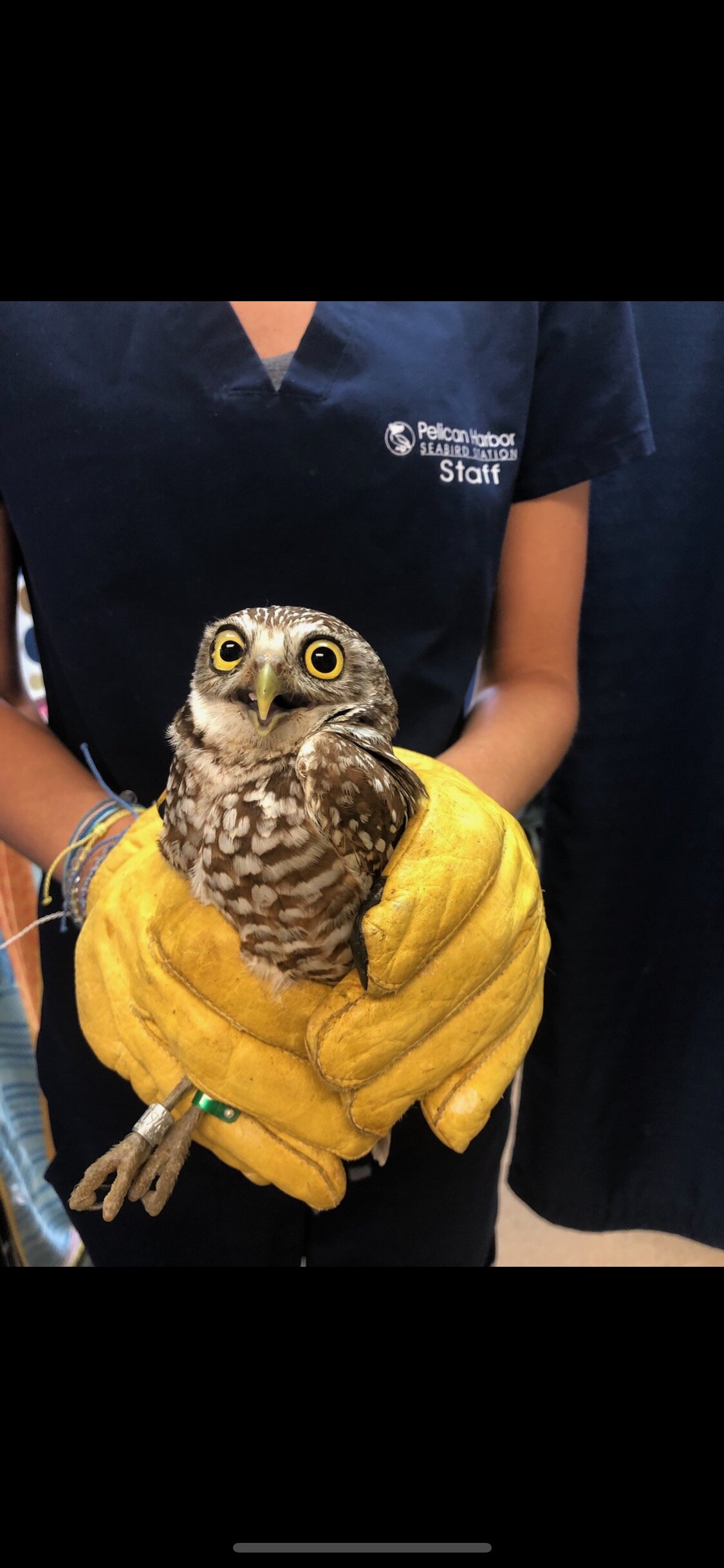
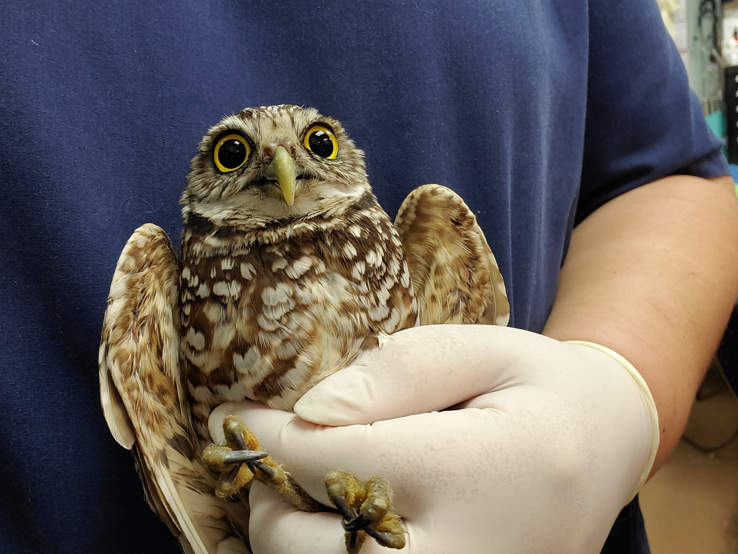
Imagine you are walking on a sunny day in the lovely city of Cape Coral and stumble across a burrow! You wonder, who made this carefully crafted burrow their home? Is it a mole? Is it a gopher? It’s a burrowing owl! With bodies reaching a maximal length of 9.8 inches and a wingspan of 21.6 inches, these one of a kind avians are one of the smallest owls in the State of Florida and are classified as State Threatened by the Florida Fish and Wildlife Conservation Commission (FWC)., These ground-dwelling owls create burrows and nests underground, as hinted at within their scientific name, Athene cunicularia – cunicularia meaning to mine or burrow in Latin. Unlike the usual image of owls peering down from trees in dense forests, burrowing owls live in vast open habitats, such as grasslands. Their burrows can stretch for lengths ranging from 6 to 10 feet, consisting of intricate tunnels weaving about 3 feet below the surface containing several bends and paths at the end of which a chamber can be found that harbors their nest., Some owls will use existing burrows or even pipes to build their burrows. The entrances to these humble abodes consist of mounds of dirt, grass, human trash articles, and are also covered intentionally by the owl with animal dung to attract insects which they can then eat. Their burrows not only serve to store away food in plentiful amounts during the period of incubation and brooding, but also provide an environment in which temperatures are better regulated and further aid in preventing the owls from dehydration during very hot days. Although, with increasing human developments and urbanization, habitat loss is an imminent and ongoing threat to these owls. In response, these owls begin to build burrows in urban environments, such as golf courses or even airports. Characteristic traits of adult burrowing owls include their dazzling yellow eyes, spindly long legs that allow them to get a better outlook from the ground-level, short tails, brown dorsal feathers with white spots and white ventral feathers with brown bar-shaped patches, distinct white eyebrows and throats, and characteristic round heads without the familiar ear tufts commonly seen in woodland owls., Nestlings on the other hand have cream-colored downy feathers that have less distinct speckles. Their diet consists mostly of insects but can also include other small birds, fish, rodents, and particularly in Florida, reptiles, such as snakes, frogs, and lizards, as, after all, raptors are carnivores.

The burrowing owl is arguably a perfect embodiment of the idiom “rare bird,” as it has so many unique attributes that set it apart from other birds in the family Strigidae. In addition to their peculiar small size and absent ear tufts, these owls are diurnal rather than nocturnal during the breeding season, thus showing activity during the daytime., Known to be very animated and sprightly, it is not uncommon to see these owls bobbing and bouncing up and down, while nestlings will even playfully leap at one another and prey, accruing at the same time important behavioral skills to prepare them for hunting later in life. Interestingly, nestlings also are known to imitate the rattling of a rattlesnake from within the protection of their burrows as a defense mechanism to ward off unwanted visitors, such as predators and humans., As a consequence of being the only species that perch on the ground, burrowing owls behave differently than one would expect; for example, if bothered or hunting they can be seen pushing themselves flat against the ground or running, respectively, as opposed to flying like owls usually do. Even more fascinating is that unlike other raptors, where the female is visibly larger than the male, which is termed “reverse sexual dimorphism,” female and male burrowing owls are the same size, with the latter being only a mere 3% larger – a size difference imperceptible to the naked human eye at first glance. Furthermore, male and female Burrowing Owls bear the same coloration, which is interesting as sexual dimorphism dictates that female and male conspecifics have traits that differ between them, and in many birds, this usually results in males exhibiting a visibly larger size or significantly more extravagant plumage coloration or ornamentation– traits that serve in courtship or competition for mates. Burrowing Owls pair for life, and unusual for an owl, are not solitary, but rather prefer to live in small colonies. This can be explained by the principle of group living, believed to have evolved independently in many different species. Group living confers advantages such as greater protection against predators, group defense, increased feeding efficiency, sharing of behaviors and communicating information, increased rate of reproduction due to access to potential partners, shelter, division of labor, and social thermoregulation. Burrowing Owls exhibit such social behaviors as the male and female even take turns in incubating the eggs, foraging, and caring for the offspring. Therefore, smaller species are often found living together in groups. Lastly, due to the significant amount of time they spend underground where there is less oxygenated air, they have an adaptation that confers them an increased tolerance for carbon dioxide.

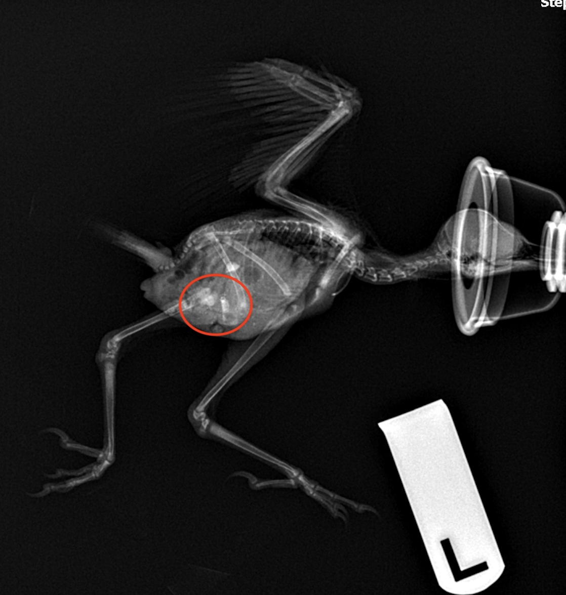
These incredible avians are, however, under threat of habitat loss, degradation, and fragmentation, as well as climate change, and rodenticide poisoning, which all have long-lasting impacts on entire ecosystems. Burrowing owls fall victim to secondary rodenticide toxicosis called “bioaccumulation” when they consume rodents that have consumed rodenticide., Second-generation anticoagulant rodenticides (SGARs), e.g., brodifacoum, are slow-acting anticoagulants, which result in the rodents not dying instantly. This leads to poisoned rodents slowly becoming weak, but because they are not instantly killed, they roam about, becoming more easily caught by predators such as Burrowing Owls. Bioaccumulation, however, causes for the period the rodent is alive to allow for a buildup of toxins to a much greater lethal dose. As the owl consumes the rodent, the toxin moves through the food web from rodents (secondary consumers) to burrowing owls (tertiary consumers), running the risk of accumulating even greater lethal amounts of poison over time at a rate greater than it can be broken down, this time in the burrowing owl. Brodifacoum is according to one source, the most widely used rat poison in the United States, and has a very long half-life in animals. A study conducted from 2006 to 2010 at Tufts Wildlife Clinic at Tufts University’s Cummings School of Veterinary Medicine reported that from 161 birds, 86% had anticoagulant rodenticide residues in their liver tissues, of which 99% had brodifacoum in their liver tissues. Moreover, only 9 of these birds that tested positive exhibited clinical symptoms, demonstrating that many affected may not show visible signs of poisoning, which is critical given the concern of bioaccumulation at an ecological level. SGARs function by inhibiting the enzyme vitamin K reductase, which under normal conditions allows for the reactivation of vitamin K – a fat-soluble vitamin that plays a vital role in the coagulation cascade by producing coagulation factors I, II, VII, IX, and X in the liver and synthesizing Protein C, Protein S, and Protein Z that serve to prevent thrombosis., During the coagulation cascade, the factors produced by vitamin K are crucial to allowing the downstream activation of prothrombin to form thrombin, the latter of which ultimately stimulates the conversion of fibrinogen to fibrin, which traps platelets to form a spongy mass that hardens into a blood clot and prevents one from bleeding out. Additionally, the Cornell Wildlife Health Lab explains that animals commonly have stores of clotting factors that delay the full effect of poisoning for 3-5 days following ingestion and the hemorrhaging onset. While coagulation panels (a blood test) exist for diagnosing anticoagulant rodenticide toxicosis in domestic animals, there are currently no existing blood tests for birds to screen for rodenticide toxicosis. Burrowing owls that get a lethal dose through consuming an affected rodent face an inhumane slow death from internal or external bleeding that causes them to weaken, which may cause them to obtain injuries in the meantime, until either the hemorrhaging or other life-threatening injuries sustained while weakened eventually kill them. If rodenticide toxicosis is evident, vitamin K can be administered to act as an antidote and help restore the blood coagulation cascade to normalcy. Additionally, stomach flushing, inducing vomiting, and administering activated charcoal are all treatments that may be used to prevent additional absorption of toxin by the body. 

Raptors such as Burrowing Owls play a critical ecological role both in the wild and in urban areas. Rodenticide toxicosis in these incredible avians is not just an issue to be left to conservationists and wildlife rescue and rehabilitators, but is a public issue that must be addressed at the source to prevent further systemic damage throughout food webs, as maintaining a sustainable and biodiverse ecosystem is a necessary goal for conserving, protecting, and restoring healthy and vibrant wildlife and their habitats and by extension and directly in relation also human health, as we are all connected and a part of the planet’s ecosystem.

Sources:

Conway, Courtney J. “Burrowing Owls: Happy Urbanite or Disgruntled Tenant?” In Urban Raptors: Ecology and Conservation of Birds of Prey in Cities, edited by Boal C.W., and Dykstra C.R., Washington, DC: Island Press, 2018. 

Cornell University. “Burrowing Owl: Identification.” The Cornell Lab of Ornithology

Cornell University. “Raptors and Rat Poison.” The Cornell Lab of Ornithology

Cornell University. “Rodenticide Toxicity.” Cornell Wildlife Health Lab

Florida Fish and Wildlife Conservation Commission. “Burrowing Owl.”

Khan, Safdar A., and Mary M. Schell. “Anticoagulant Rodenticides (Warfarin and Congeners).” Merck Veterinary Manual. Last modified October, 2014.

Libre Texts. “Role of Vitamin K.” Last modified August 13, 2020.

Murray, Maureen. “Anticoagulant Rodenticide Exposure And Toxicosis In Four Species Of Birds Of Prey Presented To A Wildlife Clinic In Massachusetts,” 2006-2010. Journal of Zoo and Wildlife Medicine 42, no. 1 (2011): 88–97.

National Audubon Society. “Guide to North American Birds: Burrowing Owl.”

National Geographic. “Biomagnification and Bioaccumulation.”

National Marine Sanctuary Foundation. “Sea Wonder: Burrowing Owl.”

Owl Research Institute. “Burrowing Owl: Athene cunicularia.”

Runnells, Charles. “Cape Coral's burrowing owls: Everything you need to know about the city's famous bird.” The News-Press. February 23, 2019.  


Vallejo, David. “Animal’s social behavior: Group living.” Zoo Portraits. July 17, 2018.

A Blue Jay's Last Chance

Written by: Daniela Hojda, PHSS Intern

Pelican Harbor sees Blue jay nestlings all the time, so when patient 21-1193 arrived at Pelican Harbor cold, depressed, and dehydrated, the rehab team knew exactly what to do to save it. It was immediately placed in an incubator to try to bring its body temperature up, and given several medications to help with each issue it presented with. Patient 1193 was given subcutaneous fluids with vitamin B complex, which are vital in helping resolve a patient’s dehydration. Often, vitamin B complex is added to the subcutaneous fluids to increase a patient’s appetite and to increase their metabolic and brain function. Also, one of the staff members heard crackles when she listened to the patient’s lungs, indicating that 1193 had aspirated. Aspiration is usually seen in baby birds when they are not brought into Pelican Harbor immediately after being found. Before bringing a baby bird to Pelican Harbor, people sometimes try to take care of and feed it. It is very easy for baby birds to aspirate food if not fed correctly, and this may lead to several health problems such as aspiration pneumonia, which in such tiny patients, is often fatal. Patient 1193 was prescribed sulfadiazine/trimethoprim (SDT), a broad spectrum antimicrobial medication, in order to help resolve the aspiration. After a few hours at Pelican Harbor, patient 1193 was much more alert and active. By the next day it seemed like a perfectly healthy nestling.

Unfortunately, two days after patient 1193 was brought to Pelican Harbor, the rehabilitation staff noticed that its right leg was resting in a concerning position, and the patient struggled to stand on it. The staff took radiographs to try and figure out why patient 1193 could not bear weight properly on its right leg. They found an old fracture of the right tibiotarsus, which had healed completely unaligned. Because the fracture was not aligned before healing, the patient’s leg was permanently bent in a way that made it unable to stand properly. This was a very worrying finding for patient 1193’s future, because an animal that is unable to use one of its legs cannot be released back into the wild. The decision was made to give patient 1193 a fighting chance by performing a surgery where the veterinarian will realign the old fracture so it can heal correctly. Once the bone is realigned, the veterinarian will then put a pin through the bone to hold the bone in place and keep the broken ends aligned. Although this surgery is extremely risky for such a small and young bird, this was patient 1193’s last chance at recovery. The veterinarian used X-rays taken before the surgery so she could get a good look at the bone that needed to be realigned. After the surgery, the staff took another set of X-rays to determine if the pin was in the right place for the bone to heal correctly. The placement of the pin looked promising, and if the patient survived the recovery period after anesthesia, the prognosis for patient 1193 was positive but still guarded. However, it’s important to remember that surgery on such a small and fragile animal is often extremely stressful which makes the recovery process extremely challenging.

Although everyone at Pelican Harbor had high hopes for patient 1193, unfortunately the stress of the surgery and anesthesia took a toll and the patient died a few hours after surgery. The staff at Pelican Harbor gave patient 1193 a fighting chance with this surgery; however, the surgery proved to be too stressful for the young Blue Jay. This type of surgery is one of the procedures used to align fractures in birds; however, the big challenge for this particular patient was how small and young it was, making it a much more risky surgery. If the patient had survived the surgery, the pin would have remained in the bone for a little over a week until a callus around the fracture formed. The patient would then be sedated again to remove the pin. Once the pin is removed we would start extensive physical therapy to ensure the patient has full use of the leg. If the patient maintained full use of the leg and no other issues arose then the patient would be a candidate for release. It’s important to remember that in wildlife medicine, rehabbers must consider that once an animal is released back into their habitat, that animal must be able to fend for themselves. An animal that has any health issues or anatomical flaw that reduces their fitness will most likely not survive in the wild.